Seamless Healing: Color-Reveal Nano Wound Dressings- NO for Unwrapping!
Dr. Danushika Manathunga -Senior Lecturer , Department of Biosystems Technology, Faculty of Technology, University of Sri Jayewardenepura

Wound management represents a significant global challenge, imposing a significant financial burden on the healthcare system. This is primarily due to the rapid growth of chronic diseases such as diabetes, obesity, and the aging population. The ability to detect pathogenic infections and immediate administration of the drugs at the wound site is of the utmost importance to expedient patient care. Herein, this article focuses on the recent work conducted concerning the development of color-changing wound dressings that change color in response to stimuli, particularly pH. It further explores the information on the use of both synthetic and natural dyes for this purpose, emphasizing the importance of promoting natural, biocompatible dye molecules to align with sustainable concepts. The discussion then extends to the novel advancements in developing multi-functional color-changing dressing materials, especially the possibility of digital image capturing via smartphones which serves as a means for record and sharing wound conditions with healthcare professionals for remote assessment and treatment guidance. The work presented here will give a better understanding of how to use color-changing dressings to eliminate the need for the unwrapping process to assess the wound healing status, an otherwise unpleasant experience for the patient. The proposed technology holds great promise in managing chronic and acute injuries resulting from trauma, surgery, or diabetes.
Key Words: Color, pH, wound dressings, dyes, chronic, acute, wounds
Content
The skin being the largest organ of the body acts as the first line of physical defense mechanism over pathogen invasion, involved in regulating the electrolyte balance and temperature control. However, due to the thin and delicate nature of the skin, it is more labile to get damaged due to acute trauma, severe burns, multiple disease conditions such as ulcers, and diabetes that may lead to severe wounds1.
The skin generally possesses its’ natural self-regeneration potential leading to wound healing. Wound healing is a complex biochemical process consisting of four main stages such as homeostasis (blood clotting), inflammation, tissue growth and tissue regeneration or maturation. During the blood clotting that takes place a few minutes after the injury, the platelets arrange themselves one after the other where then in contact with collagen leading to the generation of an amorphous structure. This activates the release of thrombin and the coagulation process with the participation of the fibrin that generates a mesh-like structure as indicated in Figure 1. This prevents the bleeding and allows the inflammatory reactions to get activated. During the inflammation, the major white blood cells such as macrophages are involved in phagocyting the dead cells, infectious bacteria, and debris. In addition, they are also involved in the release of cytokines that promote the reconstruction of the tissue with the help of fibroblast and angiogenesis. Hereafter the extracellular matrix (ECM) formation via collagen, epithelialization leads to wound remodeling. Hereafter the excess collagen removal and the wound contraction would allow the scar to build up the required strength1.
Depending on the way how the wound is getting healed, the wounds can be categorized in to main two categories namely acute and the chronic wounds. Among the two types, acute wounds are typically the wounds that are healed completely within 8-12 weeks ending up with minute scarring. These wounds occur mainly due to the mechanical injuries. On the other hand, the acute wounds arise due to severe chemical, radiation and thermal burns which extends the healing process more than 12 weeks associated with prolonged infections. This delayed healing could be a result of a secondary medical comorbidies such as diabetes, cancers and weakened angiogenesis2. This has become a serious medical challenge leading to long hospital stays, septic shock, multiorgan disorder leading to increased number of deaths in recent years3. In worldwide around 10% of mortality and 12% of morbidity is annually reported in each year due to wound injuries4.
Current practices on managing the wounds include the use of wound dressing materials that could ease the pain, prevent further progression of pathogenic infections, minimize further injury, allow the moisture management around the wounds, absorb the excess exudate and provide an ideal environment to promote the wound healing process3. The key characteristics of an ideal wound dressing is listed in the Table 1. Nevertheless, the use of wound dressings is associated with a long history, where people have used various natural leaves, ointments and cloth to cover the wounds and to reduce the pain. Until now numerous wound dressing materials have been developed in clinical setting where their application has been dependent on the type of the injury. These materials include traditional dressings like gauzes, cotton wools as well as much developed materials like hydrogels and foams. These are dedicated to deliver antimicrobials, regenerative medicines and other bioactive compounds. These wound dressings play a pivotal role in facilitating the interaction of medications, growth factors with the regenerating cells and extracellular matrix. Among different wound dressings the silver-impregnated dressings such as Acticoat (Smith & Nephew), Fibracol (Johnson & Johnson), and Silvasorb (Medline) have got special attention over the years due to their distinct ability to prevent infections3,5,6.
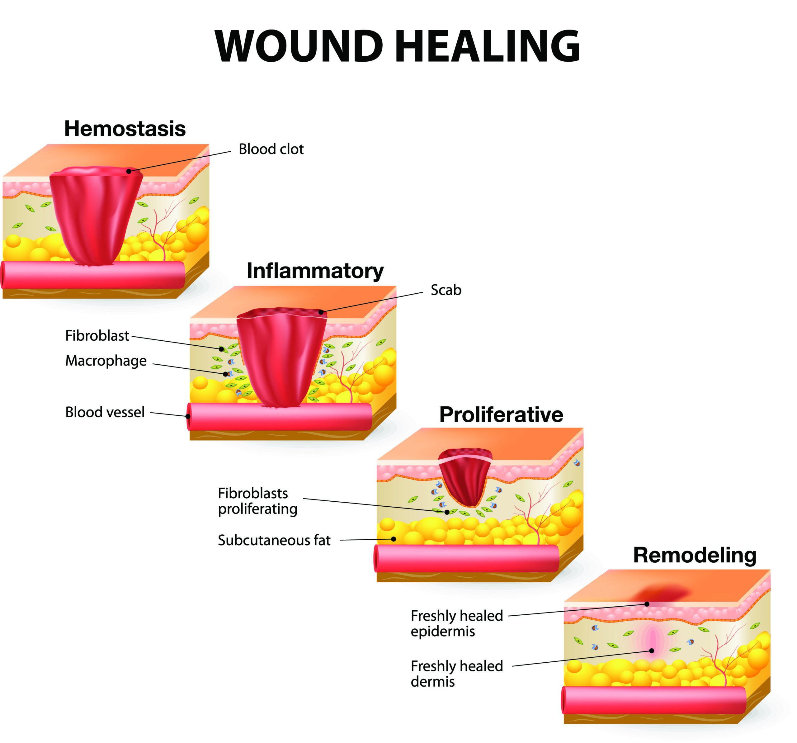
Figure 1: Stages in wound healing process (Source: https://www.shieldhealthcare.com/community/popular/2015/12/18/how-wounds-heal-the-4-main-phases-of-wound-healing/ accessed 02/03/2024)
Table 1. Key characteristics expected in an ideal wound dressing3.
| Feature
|
Description |
| Absence of toxic substances | Not inducing any toxicity or damage to the growing cells. |
| Minimize the bacterial infection | Prevent the bacterial invasion that leads to delay of wound healing |
| Adhesiveness | Provide the required adhesive property to hold the content |
| Moisture content | Maintain optimal moisture content to facilitate the cell migration and proliferation |
| Thermal insulation | Maintain the optimal temperature in the wound site. |
| Exudate management | Absorb excess wound exudate and minimize leaking, unpleasant appearance |
| Oxygen permeability | Accelerate the oxygen diffusion and supply to the wound bed to facilitate the cell proliferation. |
| Mechanical property | Provide the required strength to the regenerating wound |
| Minimal pain | Minimize the pain during the application and removal of the dressing |
| Cost effectiveness | Be affordable |
| Availability | Available for majority of patients |
Even though various wound dressings have been introduced into the market, several persistent challenges hinder the optimal delivery of the benefits to the patients. One of the notable issue is the requirement of change of the dressings frequently for the visual inspection of the wound healing progression that cause a discomfort to the patient. Moreover, the development of antibiotic resistance, delayed healing, lack of high-tension endurance and the inability to adjust the dynamics in the wound environment (e.g. control the release of antibiotics or the medications) are among the other challenges underscore the pressing need on developing smart multifunctional wound dressings that could simultaneously monitor the wound conditions and indicating on proper treatment option at the same time3,5,6. This will allow the user to identify the optimal wound dressing type with the required elements for the quick facilitation of the wound healing process7.
Among the said points the frequent dressing changes for the visual examination of the status of the wound is a critical factor that guide the clinicians to take the next decisions about the wound progression. This will not only inflict trauma to the patient, but contributes for an overall unforgettable bad experience to the wearer/ patient. More this practice potentially possess the risk of introducing new disease causing infectious agents triggering the wound infections8.
To address this issue, over the last few years attempts have been taken to generate smart wound dressings with in-built sensors that could capture the status of the wound with the help of the change of a biomarker or wound parameter (pH, temperature, specific enzymes, glucose level, moisture level, oxygen concentration) present in the wound site, where the wearer will be notified with the change of a physical parameter such as color, heat etc. This has allowed the real time monitoring of the wound status without unwrapping, coupled with the time-controlled release of drug molecules to the wound and the tissue regeneration with the application of the same wound dressing material5.
Among many biomarkers, the change of the pH in the wound site is a found to be a clear alternative indication of the wound healing process as the different stages of the wound healing process is clearly associated with definite pH regimes. In this regard, wound healing is experimented to be approaching to a low pH while the high pH values allow the wound to be persistent for a certain period of time. The pH of these prolong/ chronic wounds are found to be in the pH range of 7.15-8.9. Therefore, the gradual transition of the alkaline pH in to neutral to acidic pH range is considered as a good indication about the healing process. Hence, the monitoring of the pH will be a good strategy to determine the status of the wound without the requirement for unwrapping6.
The indication about the pH change for it to be diagnosed visually, it is required to be coupled with a readily identifiable color change. For this purpose, a halochromic dyes can be utilized owing to their pH sensitive chromophores which could lead to the generation of vibrant colors. Considering this concept, numerous efforts have been made to generate pH sensitive color indicative wound dressing materials. Among various materials developed the use of pH sensitive hydrogels9, textile materials10, electrospun nanofibers11 have received increasing attention over the years. These materials convey the exact information of the wound with the facilitation of a color change that could be easily identified by the naked eye.
Furthermore, current work also suggests the possibility of attaining a quantitative continuous measurement of the wound pH by coupling it to smart devices such as smart phones which could capture high quality images, processed via specific software to generate a value, which helps the clinicians to get a direct understanding about the wound status3.
Among different types of wound dressing materials, hydrogels have become much familiar in the clinical setting as they have a special ability to mimic the environment of the human skin. Various hydrogel types such as a films, sprayable hydrogels and injectable gels have been designed with inbuilt sensors to assess the wound pH2. These hydrogels have been prepared using natural biocompatible polymers such as gelatine, collagen, alginate, chitosan, hyaluronic acid as well as using synthetic polymers like poly [lactic-co-glycolic acid] PLGA and poly(vinyl alcohol) PVA. Furthermore, attempts have been also made to prepare composite hydrogels consisted of both synthetic and natural polymers to obtain advanced wound healing properties5. Some of these studies have also focused on the development of color changing multifunctional wound dressings where it could release some antibiotics in pulsative manner. One such commercially launched product, is “GelDerm” that has been invented by the Prof. Akbari the research team (Figure. 2). This hydrogel is capable of continuously releasing the antibiotic agent at the wound site once the dressing is placed on the wound. Compared with the conventional approaches, this gel is specifically capable of generating a colorful map of the wound site with the variation of its pH, in addition to its ability to continuously release the antibiotics and the moisture management.
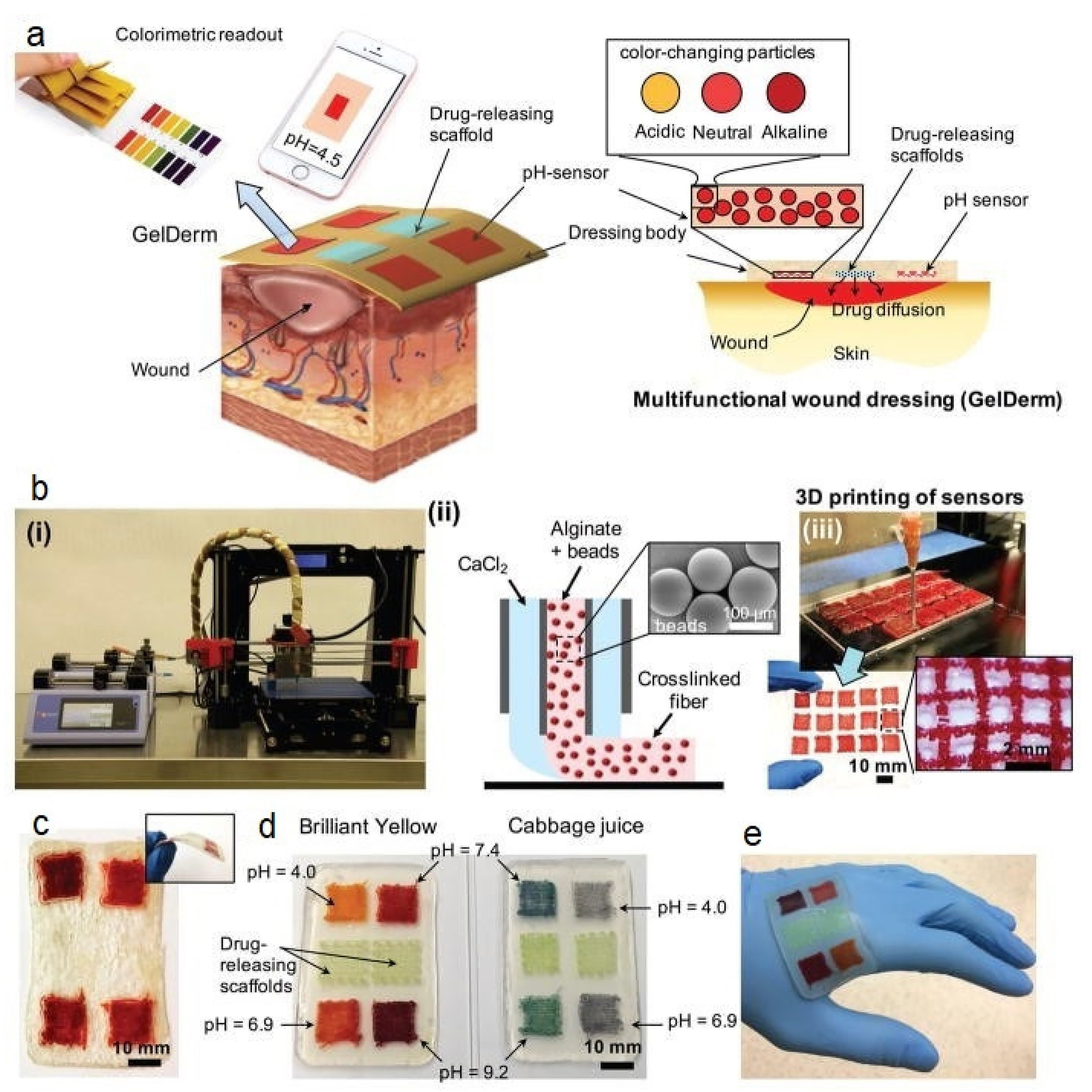
Figure 2. An integrated system of GelDerm with pH sensitive dye (Cabbage extract) that has been developed to measure the wound healing process. A) Schematic representation of GelDerm treatment of epidermal wounds, with pH-sensitive and drug-eluting components. B-i) Porous sensors were fabricated using a 3D bioprinter equipped with a co-axial flow microfluidic nozzle (i). B-ii) Schematic of fiber deposition using the co-axial flow system. B-iii) 3D printer can be programmed to produce arrays of porous sensors for fabrication of large-scale dressings. C) Dressings can be lyophilized and sterilized for storage and transportation. D) Synthetic Brilliant Yellow and naturally derived cabbage juice were used as model pH indicators for the fabrication of the sensors (Reprinted with the permission of Akbari et al., 20283).
As indicated in Figure 3., beyond hydrogels, pH sensitive textile materials, cotton swabs and mesoporous alginate beads impregnated with halochromic dyes have been also developed to cater the said purpose.12 Notably recent attention has been also devoted on exploring the use of electrospun nanofibers as wound healing materials. These materials are known to have pronounced compatibility with the extracellular matrix (ECM), accounting with high surface area to volume ratio promoting oxygen permeability and exudate absorbing capacity. They have been also tailored to bear some additional characteristics such as the release of antibiotics and antioxidant compounds that facilitate the wound healing process13.
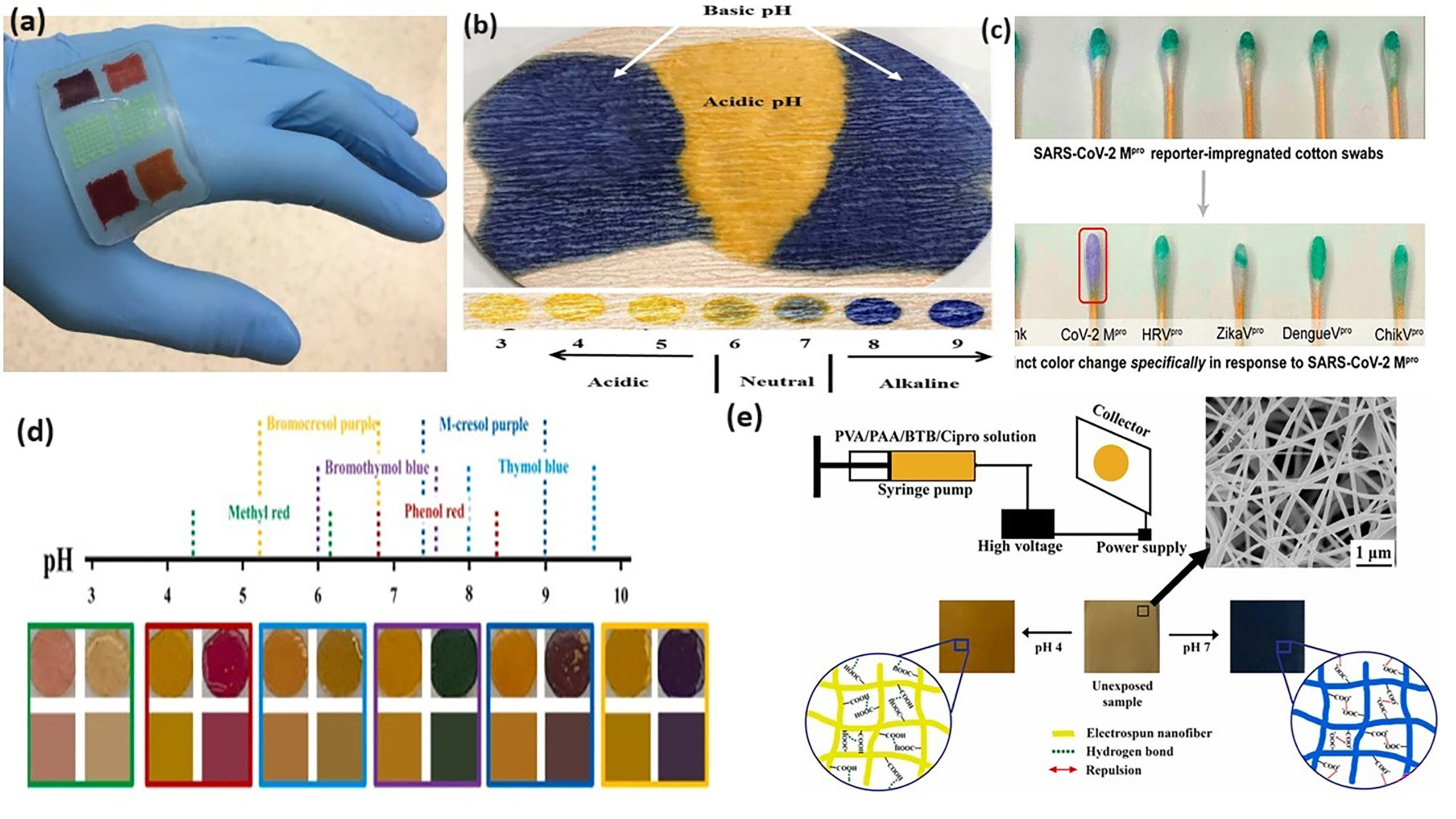
Figure 3. Different types of pH sensitive color changing wound dressing substates (a) hydrogels, (b) textiles, (c) cotton swabs, (d) mesoporous silica beads and (e) electrospun nanofibers(Reprinted with the permission from https://www.chemistryviews.org/details/news/10644597/Multifunctional_Hydrogel_Wound_Dressing/, Hu et al., 202014, Mohr at al., 201715, Yu et al., 202216 and Arafat et al., 202111)
In these smart wound dressings, the key factor that determines the indication about the wound status as well as the parameter that defines the rate at which the wound healing takes place is the pH of the wound which is detected via a pH sensitive indicator dye containing chromophores. Up to now various halochromic dye molecules such as bromocresol green, bromocresol purple17,18, methylene blue19 and phenol red20 have been used to fulfill the said purpose where they have exhibited a clear color demarcation between the pH of a wound and it’s healed status. In addition to those the fluorescent pH sensing dyes have been also popular in making pH sensitive wound dressings21. However, it has been noticed that the use of synthetic halochromic dyes is associated with some toxicity and biocompatibility issues and therefore found to be not suitable for medical applications. In addition to it has been also noticed that their pH response is restricted to a particular range and therefore demand for the presence of combination of dyes to provide a better response22.
Therefore, recent interest has been concentrated on the use of natural pigments such as anthocyanin, curcumin, betalain, chlorophylls and betacyanins for the development of color changing wound dressings. These dyes have been extracted from different plant sources and plant parts. Some of these were also found to contain antioxidant, anti- inflammatory, anti-microbial and anti-cancer properties. Among these dye materials, anthocyanins have been much popular because of their ability to extend the color response over a wide color spectrum where the pH falls over the range of 1-14. This color variation is clearly from the pink (acidic) to, purple, blue to green (basic) hue with the change of the pH from acidic to basic as indicated in Figure 4. Anthocyanins are widely extracted from sources such as berries, currants, grapes and some tropical fruits23. Moreover they are also found to be rich in some flowers such as Hibiscus and Butterfly pea24,25.
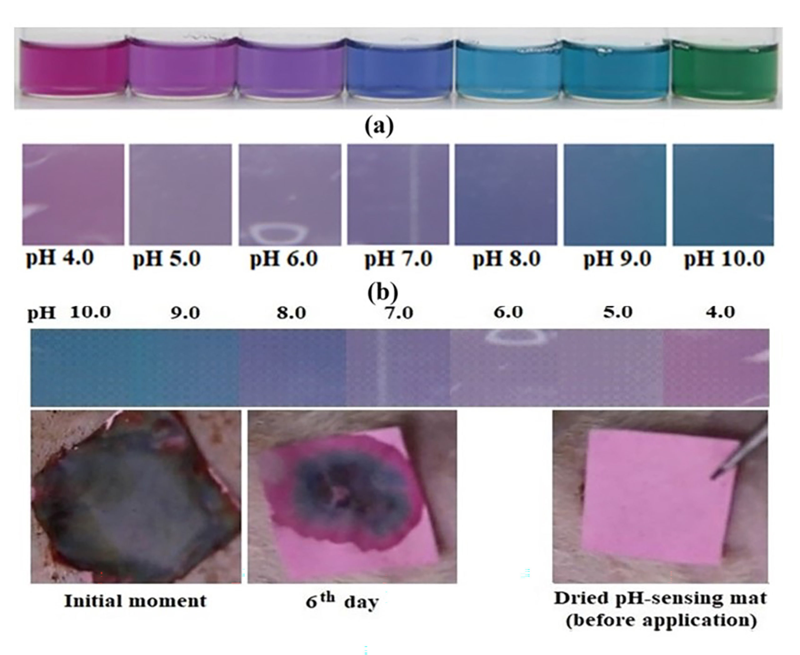
Figure 4. Color response of anthocyanin dye over the pH range of 4-10 and the application of anthocyanin impregnated nanosheet on an open wound to measure the wound pH (Reprinted with the permission from Karaca at al., 20216).
Given below in Figure 5., are some of the wound dressings materials that have been prepared with the incorporation of natural dyes and their respective color changing behavior. They have been mainly prepared by utilizing the materials that can be commonly found in nature, promoting the sustainability concept.
Considering the reason scientific advancements, breakthroughs and the commercialization aspects with respect to the development of novel wound dressing materials, following links will provide you with the most current work that have been conducted in this area of research.
https://newatlas.com/color-changing-dressing/16808/
https://www.trendhunter.com/trends/colorchanging-wound-dressings
https://insights.globalspec.com/article/20383/color-changing-wound-dressing-reveals-infections
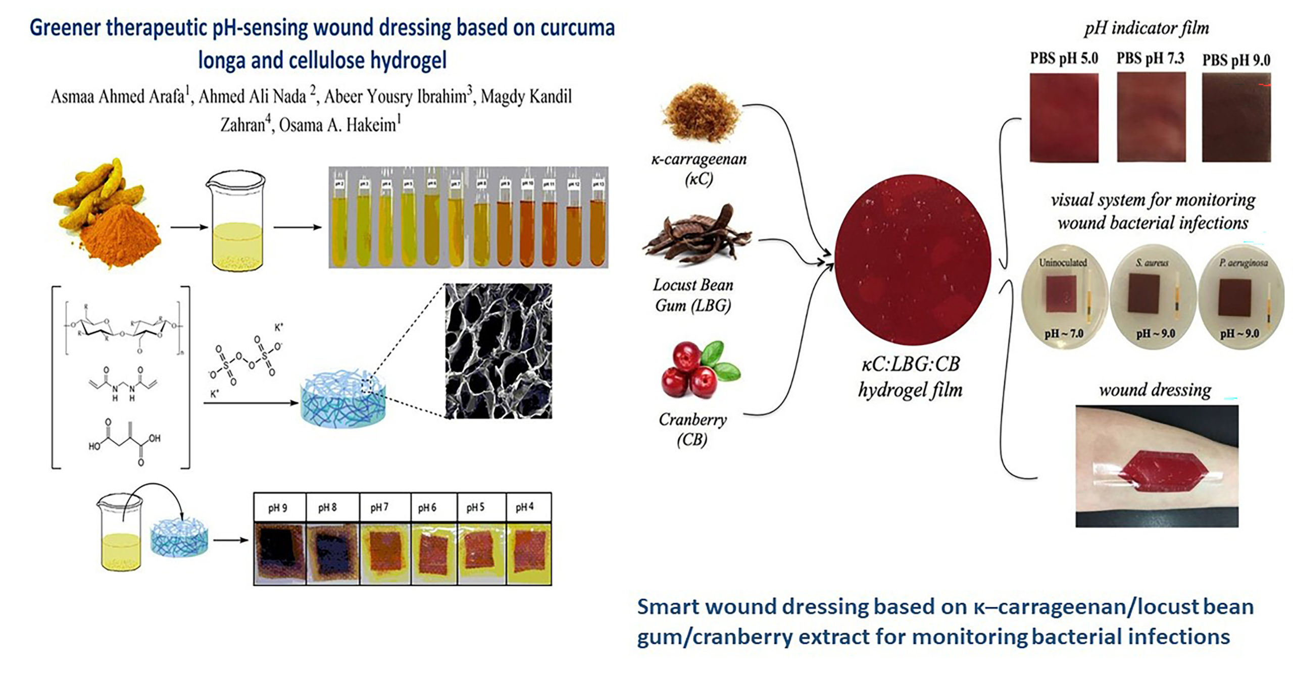
Figure 5. Curcumin (turmeric extract) and cranberry extract incorporated smart dressings with color changing ability (Reprinted with the permission from Arafa et al., 20219 and Zepon et al., 201926).
As a conclusion, it can be noted that the application of these materials in large scale for clinical settings, have not been achieved at the present status, highlighting some challenges that are yet to be overcome for its successful implementation. They will include the sophisticated layout of the materials negatively affecting the scalability, shortage of natural supply of dyes, lack of biocompatibility studies and the fear related to the use of nanomaterials as well as poor effectiveness when targeted to provide multifunctional ability. However, irrespective of the mentioned hurdles the next generation wound healing with color changing pH sensitive property will most likely to focus in the development of cell-laden: smart with nanomaterials with integrated sensors to detect and to treat non-healing wounds.
References
- Pan, N., Qin, J., Feng, P., Li, Z. & Song, B. Color-changing smart fibrous materials for naked eye real-time monitoring of wound pH. J. Mater. Chem. B 7, 2626–2633 (2019).
- Tavakoli, S. & Klar, A. S. Advanced hydrogels as wound dressings. Biomolecules 10, 1–20 (2020).
- Bahram Mirani. An Advanced Multifunctional Hydrogel-Based Dressing for Wound Monitoring and Drug Delivery. Adv Heal. Mater. 6, 1–26 (2017).
- Chowdhury, S. & Chakraborty, P. pratim. Universal health coverage ‑ There is more to it than meets the eye. J. Fam. Med. Prim. Care 6, 169–170 (2017).
- Li, M. et al. Smart and versatile biomaterials for cutaneous wound healing. Biomater. Res. 27, 1–32 (2023).
- Pakolpakçıl, A. et al. Design and in vivo evaluation of alginate-based pH-sensing electrospun wound dressing containing anthocyanins. J. Polym. Res. 28, 1–13 (2021).
- Dabiri, G., Damstetter, E. & Phillips, T. Choosing a Wound Dressing Based on Common Wound Characteristics. 5, 32–41 (2016).
- Gefen, A. et al. How Should Clinical Wound Care and Management Translate to Effective Engineering Standard Testing Requirements from Foam Dressings? Mapping the Existing Gaps and Needs. Adv. Wound Care 13, 34–52 (2024).
- Arafa, A. A., Nada, A. A., Ibrahim, A. Y., Zahran, M. K. & Hakeim, O. A. Greener therapeutic pH-sensing wound dressing based on Curcuma Longa and cellulose hydrogel. Eur. Polym. J. 159, 110744 (2021).
- Gamerith, C. et al. pH-responsive materials for optical monitoring of wound status. Sensors Actuators, B Chem. 301, 126966 (2019).
- Arafat, M. T., Mahmud, M. M., Wong, S. Y. & Li, X. PVA/PAA based electrospun nanofibers with pH-responsive color change using bromothymol blue and on-demand ciprofloxacin release properties. J. Drug Deliv. Sci. Technol. 61, (2021).
- Eskilson, O. et al. Nanocellulose composite wound dressings for real-time pH wound monitoring. Mater. Today Bio 19, (2023).
- Han, Z. et al. pH-Responsive wound dressings: advances and prospects. Nanoscale Horizons 8, 422–440 (2023).
- Vu, H. et al. A Device to Predict Short-Term Healing Outcome of Chronic Wounds. Adv. Wound Care 9, 312–324 (2020).
- Schaude, C. et al. The development of indicator cotton swabs for the detection of pH in wounds. Sensors (Switzerland) 17, (2017).
- Patel, S. et al. Wearable electronics for skin wound monitoring and healing. Soft Sci. 2, (2022).
- van der Schueren, L. & de Clerck, K. Coloration and application of pH-sensitive dyes on textile materials. Color. Technol. 128, 82–90 (2012).
- Kurečič, M. et al. A multifunctional electrospun and dual nano-carrier biobased system for simultaneous detection of pH in the wound bed and controlled release of benzocaine. Cellulose 25, 7277–7297 (2018).
- Li, Z. et al. Silk fibroin–gelatin photo-crosslinked 3D-bioprinted hydrogel with MOF-methylene blue nanoparticles for infected wound healing. Int. J. Bioprinting 9, (2023).
- Dressings, W. A pH-Indicating Colorimetric Tough Hydrogel Patch Wound Dressings. (2017) doi:10.3390/polym9110558.
- Al-Hawat, M. L. et al. Fluorescent pH-sensing bandage for point-of-care wound diagnostics. Aggregate 1–8 (2023) doi:10.1002/agt2.472.
- Adamu, B. F. et al. Self-Responsive Electrospun Nanofibers Wound Dressings: The Future of Wound Care. Adv. Mater. Sci. Eng. 2022, (2022).
- Tena, N., Martín, J. & Asuero, A. G. State of the art of anthocyanins: Antioxidant activity, sources, bioavailability, and therapeutic effect in human health. Antioxidants 9, (2020).
- Salacheep, S. et al. Optimization of ultrasound-assisted extraction of anthocyanins and bioactive compounds from butterfly pea petals using Taguchi method and Grey relational analysis. J. Food Sci. Technol. 57, 3720–3730 (2020).
- Maciel, L. G. et al. Hibiscus sabdariffa anthocyanins-rich extract: Chemical stability, in vitro antioxidant and antiproliferative activities. Food Chem. Toxicol. 113, 187–197 (2018).
- Zepon, K. M. et al. Smart wound dressing based on κ–carrageenan/locust bean gum/cranberry extract for monitoring bacterial infections. Carbohydr. Polym. 206, 362–370 (2019).
Dr. Danushika Manathunga
Senior Lecturer
Department of Biosystems Technology, Faculty of Technology, University of Sri Jayewardenepura